Atypical Bacterial Pneumonia Imaging: Overview, Radiography, Computed Tomography
Pneumonia is predominantly a clinical syndrome.The classic etiologic agents of atypical pneumonia are Legionella species, Mycoplasma pneumoniae, and Chlamydia pneumoniae. Many other diseases, caused by various pathogens, should be considered in the differential diagnosis. Such etiologic agents include fungi, mycobacteria, parasites, and viruses (eg, influenza virus, adenovirus, respiratory syncytial virus, human parainfluenza virus, measles, varicella zoster, Hantavirus).
In immunosuppressed patients, outbreaks of isolated cases of respiratory virus infections with atypical presentations are reported. These infections can be severe and may have concomitant bacterial etiologies. In endemic areas, certain zoonotic infections should be considered when patients present with atypical pneumonia. Noninfectious etiologies must be considered in atypical and nonresolving pneumonias.
During the latter half of the 19th century, by which time physicians had embraced autopsy as an essential learning tool, pneumonia diagnoses were usually made post mortem. With the discovery of x-rays (1895), chest radiography became part of the routine evaluation of pneumonia in patients with suggestive signs and symptoms. Patients who presented with fever, shaking chills, and rust-colored sputum (which under examination showed gram-positive diplococci in chains) and whose chest radiographic findings were suggestive of pulmonary infection were considered to have typical pneumonia. Scans of atypical bacterial pneumonia are depicted below.
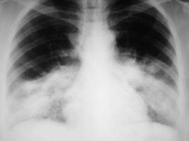 A 53-year-old patient with severe Legionellapneumonia. Chest radiograph shows dense consolidation in both lower lobes.
 A 40-year-old patient with Chlamydia pneumonia. Chest radiograph shows multifocal, patchy consolidation in the right upper, middle, and lower lobes.
 A 38-year-old patient with Mycoplasma pneumonia. Chest radiograph shows a vague, ill-defined opacity in the left lower lobe.
 Chest computed tomography scan shows ill-defined, airspace infiltrate in the left lower lobe.
A phase of active hyperemia occurs, lasting approximately 24 hours before radiologic consolidation of the alveoli appears. This phase is characterized by engorgement of the arterial blood vessels. Edematous fluid, which may be seen in the alveolus, contains few exudative cells.
The next stage is referred to as red hepatization. Neutrophils and fibrin material fill the alveoli, and massive extravasation of red blood cells produces a homogeneous opacity.
The red hepatization phase is then followed by gray hepatization. Fibrin and exudative cells accumulate, appearing on radiographs as a clear zone adjoining the alveolar and acinar cells.
If the process extends to the pleural space, associated empyema may be present.
The radiographic characteristics of Legionella, Mycoplasma, and Chlamydia pneumonias are discussed in this section.
Legionella species are implicated in 2-15% of community-acquired pneumonia (CAP) cases.
These organisms usually cause a patchy, localized infiltrate in the lower lobes. Associated hilar adenopathy may be present. Pleural effusion is seen in up to 30% of cases. In rare instances, Legionella infection is associated with cavitation and a masslike appearance.
Radiologic resolution of Legionella pneumonia may take 6-12 months. Permanent residual fibrosis is observed in as many as 25% of patients. An early progression of infiltrates can occur despite clinical improvement.Legionella pneumonia is depicted in the image below.
A 53-year-old patient with severe Legionellapneumonia. Chest radiograph shows dense consolidation in both lower lobes.
Chest radiographs cannot be used to distinguish nosocomial legionellosis (Legionnaires disease) from other pneumonias.
M pneumoniae is implicated in 2-30% of all cases of CAP. Mycoplasma pneumonia is usually mild and results in a rapid resolution of any radiologic findings. However, it tends to be more severe in patients with sickle cell anemia. Radiographic resolution in 40% of patients occurs in 4 weeks, and 80% of cases resolve by 8 weeks. Residual radiographic abnormalities are uncommon.
The infiltrates in Mycoplasma pneumonia can be unilateral, multilobar, or bilateral. In about 20% of patients, pleural effusion or hilar adenopathy may be present. Mycoplasma pneumonia is depicted in the image below.
A 38-year-old patient with Mycoplasma pneumonia. Chest radiograph shows a vague, ill-defined opacity in the left lower lobe.
The infiltrates may be subsegmental or more extensive in elderly patients; pleural effusions are rarely seen. Chest radiographs show 50% resolution in 4 weeks. In 20% of cases, resolution takes longer than 9 weeks. Chlamydia pneumonia is depicted in the image below.
A 40-year-old patient with Chlamydia pneumonia. Chest radiograph shows multifocal, patchy consolidation in the right upper, middle, and lower lobes.
Radiologic findings alone are not reliable in differentiating pneumonia into typical or atypical forms. Therefore, the radiographic findings described above should be used along with clinical and laboratory data to narrow the possibilities.
Structural lung disease with abnormal lung parenchyma affects the pattern of infiltrates. In cases of severe emphysematous lung disease, clinicians may tend to underestimate the presence of infiltrates on chest radiographs.
Computed tomography (CT) scans are increasingly being used in clinical practice. Various authors have questioned CT scanning's usefulness in evaluating consolidations, suggesting that the value of CT in the diagnosis of pneumonia is limited to specific cases involving the following:
Tanaka et al compared high-resolution CT (HRCT) scan findings in CAP with pathologic findings and evaluated the role of HRCT scanning in differentiating between bacterial and atypical pneumonias in 32 patients with CAP (18 with bacterial pneumonia, 14 with atypical pneumonia).
Bacterial pneumonia often resulted in airspace consolidation with a segmental distribution (72%) that typically occurred toward the middle and outer zones of the lungs. Atypical pneumonias included Mycoplasma and Chlamydia pneumonias, as well as influenza viral pneumonia. These conditions frequently caused a centrilobular shadow (64%), an acinar shadow (71%), and/or airspace consolidation (57%) and ground-glass attenuation (86%) with a lobular distribution. The lesions were often distributed to the inner, middle, and outer layers of the lung (86%). A CT scan depicting chlamydial pneumonia is shown in the image below.
 Chest computed tomography scan in a 45-year-old patient with Chlamydia pneumonia shows a right upper-lobe infiltrate.
Mild Legionella pneumonia may manifest with bilateral involvement of the lung parenchyma. Multiple segments are affected, and peripheral lung consolidation with ground-glass opacity and pleural effusion may be seen. With more severe infection, lung cavitation and bulging of the fissure have been reported. Residual lung parenchymal scarring can be found, even after the acute infection resolves.A CT scan depicting Legionella pneumonia is shown in the image below.
 Image in a 66-year-old patient with Legionella pneumonia. Chest computed tomography scan shows dense alveolar consolidations in both lower lobes.
Reittner et al examined 28 patients, identifying ground-glass attenuation in 24 (86%) and airspace consolidation in 22 (79%). In 13 of the latter 22 patients (59%), the areas of consolidation had a lobular distribution. Nodules were more common on HRCT scans (89%) than on radiographic images (50%), and in 24 of 28 patients (86%), the nodules had a predominantly centrilobular distribution on CT scans. Thickening of bronchovascular markings were more often found with CT scanning (82%) than with radiography (18%).
Coinfection with several organisms is not uncommon. Underlying parenchymal lung abnormalities usually predispose patients to pneumonia. Therefore, in patients with pneumonia, the overall clinical and radiologic picture must be considered in place of an independent, dichotomous view. (See the image below.)
 Although this patient smokes, this lesion most likely has an inflammatory etiology, given the clinical symptoms and a recent, normal CT scan. Appropriate management includes repeat CT scanning in 3 months if the lesion persists or enlarges despite clinical improvement.
Robbins and Kumar described Legionella infection in an HIV patient who was found on MRI to have a lesion of the splenium of the corpus callosum.
The literature suggests that ultrasonography can help in differentiating between consolidation and effusion. Consolidated lung tissue may appear as hypoechoic areas with blurred margins. The texture varies with the amount of aeration, being more heterogeneous with aeration and more homogeneous with dense consolidation. The literature also reports that ultrasonography may aid in the diagnosis of empyema and abscesses. However, the current authors believe that in clinical practice, ultrasonography's usefulness is limited to the identification and quantification of parapneumonic effusions. Once found, the area where an effusion occurs can be marked for subsequent diagnostic or therapeutic thoracentesis.
Shakeel Amanullah, MD Consulting Physician, Pulmonary, Critical Care, and Sleep Medicine, Lancaster General Hospital
Shakeel Amanullah, MD is a member of the following medical societies: American College of Chest Physicians
Coauthor(s)
Klaus-Dieter Lessnau, MD, FCCP Clinical Associate Professor of Medicine, New York University School of Medicine; Medical Director, Pulmonary Physiology Laboratory; Director of Research in Pulmonary Medicine, Department of Medicine, Section of Pulmonary Medicine, Lenox Hill Hospital
Klaus-Dieter Lessnau, MD, FCCP is a member of the following medical societies: American College of Chest Physicians, American College of Physicians, American Medical Association, American Thoracic Society, Society of Critical Care Medicine
David H Posner, MD Assistant Professor of Medicine, New York University School of Medicine; Assistant Chief of Pulmonary Diseases, Instructor, Intensive Care Unit, Education Coordinator for Pulmonary Fellowship, Lenox Hill Hospital
Mina Farhad, MD, PhD Clinical Instructor of Radiology, New York University School of Medicine; Head of Thoracic Imaging, Department of Radiology, Lenox Hill Hospital
Mina Farhad, MD, PhD is a member of the following medical societies: Radiological Society of North America
Specialty Editor Board
Bernard D Coombs, MB, ChB, PhD Consulting Staff, Department of Specialist Rehabilitation Services, Hutt Valley District Health Board, New Zealand
Chief Editor
Kavita Garg, MD Professor, Department of Radiology, University of Colorado School of Medicine
Kavita Garg, MD is a member of the following medical societies: American College of Radiology, American Roentgen Ray Society, Radiological Society of North America, Society of Thoracic Radiology
Satinder P Singh, MD, FCCP Professor of Radiology and Medicine, Chief of Cardiopulmonary Radiology, Director of Cardiac CT, Director of Combined Cardiopulmonary and Abdominal Radiology, Department of Radiology, University of Alabama at Birmingham School of Medicine
References
A 53-year-old patient with severe Legionellapneumonia. Chest radiograph shows dense consolidation in both lower lobes.
A 40-year-old patient with Chlamydia pneumonia. Chest radiograph shows multifocal, patchy consolidation in the right upper, middle, and lower lobes.
A 38-year-old patient with Mycoplasma pneumonia. Chest radiograph shows a vague, ill-defined opacity in the left lower lobe.
Chest computed tomography scan shows ill-defined, airspace infiltrate in the left lower lobe.
Chest computed tomography scan in a 45-year-old patient with Chlamydia pneumonia shows a right upper-lobe infiltrate.
Image in a 66-year-old patient with Legionella pneumonia. Chest computed tomography scan shows dense alveolar consolidations in both lower lobes.
Although this patient smokes, this lesion most likely has an inflammatory etiology, given the clinical symptoms and a recent, normal CT scan. Appropriate management includes repeat CT scanning in 3 months if the lesion persists or enlarges despite clinical improvement.
Overview
Pneumonia is predominantly a clinical syndrome.The classic etiologic agents of atypical pneumonia are Legionella species, Mycoplasma pneumoniae, and Chlamydia pneumoniae. Many other diseases, caused by various pathogens, should be considered in the differential diagnosis. Such etiologic agents include fungi, mycobacteria, parasites, and viruses (eg, influenza virus, adenovirus, respiratory syncytial virus, human parainfluenza virus, measles, varicella zoster, Hantavirus).
In immunosuppressed patients, outbreaks of isolated cases of respiratory virus infections with atypical presentations are reported. These infections can be severe and may have concomitant bacterial etiologies. In endemic areas, certain zoonotic infections should be considered when patients present with atypical pneumonia. Noninfectious etiologies must be considered in atypical and nonresolving pneumonias.
During the latter half of the 19th century, by which time physicians had embraced autopsy as an essential learning tool, pneumonia diagnoses were usually made post mortem. With the discovery of x-rays (1895), chest radiography became part of the routine evaluation of pneumonia in patients with suggestive signs and symptoms. Patients who presented with fever, shaking chills, and rust-colored sputum (which under examination showed gram-positive diplococci in chains) and whose chest radiographic findings were suggestive of pulmonary infection were considered to have typical pneumonia. Scans of atypical bacterial pneumonia are depicted below.
A 53-year-old patient with severe Legionellapneumonia. Chest radiograph shows dense consolidation in both lower lobes. A 40-year-old patient with Chlamydia pneumonia. Chest radiograph shows multifocal, patchy consolidation in the right upper, middle, and lower lobes. A 38-year-old patient with Mycoplasma pneumonia. Chest radiograph shows a vague, ill-defined opacity in the left lower lobe. Chest computed tomography scan shows ill-defined, airspace infiltrate in the left lower lobe. Radiologic phases
A phase of active hyperemia occurs, lasting approximately 24 hours before radiologic consolidation of the alveoli appears. This phase is characterized by engorgement of the arterial blood vessels. Edematous fluid, which may be seen in the alveolus, contains few exudative cells.
The next stage is referred to as red hepatization. Neutrophils and fibrin material fill the alveoli, and massive extravasation of red blood cells produces a homogeneous opacity.
The red hepatization phase is then followed by gray hepatization. Fibrin and exudative cells accumulate, appearing on radiographs as a clear zone adjoining the alveolar and acinar cells.
If the process extends to the pleural space, associated empyema may be present.
Radiography
The radiographic characteristics of Legionella, Mycoplasma, and Chlamydia pneumonias are discussed in this section.
Legionella pneumonia
Legionella species are implicated in 2-15% of community-acquired pneumonia (CAP) cases.
These organisms usually cause a patchy, localized infiltrate in the lower lobes. Associated hilar adenopathy may be present. Pleural effusion is seen in up to 30% of cases. In rare instances, Legionella infection is associated with cavitation and a masslike appearance.
Radiologic resolution of Legionella pneumonia may take 6-12 months. Permanent residual fibrosis is observed in as many as 25% of patients. An early progression of infiltrates can occur despite clinical improvement.Legionella pneumonia is depicted in the image below.
A 53-year-old patient with severe Legionellapneumonia. Chest radiograph shows dense consolidation in both lower lobes. Chest radiographs cannot be used to distinguish nosocomial legionellosis (Legionnaires disease) from other pneumonias.
Mycoplasma pneumonia
M pneumoniae is implicated in 2-30% of all cases of CAP. Mycoplasma pneumonia is usually mild and results in a rapid resolution of any radiologic findings. However, it tends to be more severe in patients with sickle cell anemia. Radiographic resolution in 40% of patients occurs in 4 weeks, and 80% of cases resolve by 8 weeks. Residual radiographic abnormalities are uncommon.
The infiltrates in Mycoplasma pneumonia can be unilateral, multilobar, or bilateral. In about 20% of patients, pleural effusion or hilar adenopathy may be present. Mycoplasma pneumonia is depicted in the image below.
A 38-year-old patient with Mycoplasma pneumonia. Chest radiograph shows a vague, ill-defined opacity in the left lower lobe. Chlamydia pneumonia
The infiltrates may be subsegmental or more extensive in elderly patients; pleural effusions are rarely seen. Chest radiographs show 50% resolution in 4 weeks. In 20% of cases, resolution takes longer than 9 weeks. Chlamydia pneumonia is depicted in the image below.
A 40-year-old patient with Chlamydia pneumonia. Chest radiograph shows multifocal, patchy consolidation in the right upper, middle, and lower lobes. Degree of confidence
Radiologic findings alone are not reliable in differentiating pneumonia into typical or atypical forms. Therefore, the radiographic findings described above should be used along with clinical and laboratory data to narrow the possibilities.
Structural lung disease with abnormal lung parenchyma affects the pattern of infiltrates. In cases of severe emphysematous lung disease, clinicians may tend to underestimate the presence of infiltrates on chest radiographs.
Computed Tomography
Computed tomography (CT) scans are increasingly being used in clinical practice. Various authors have questioned CT scanning's usefulness in evaluating consolidations, suggesting that the value of CT in the diagnosis of pneumonia is limited to specific cases involving the following:
- An indistinct, abnormal opacity on chest radiographs
- Patchy, ground-glass, linear, or reticular opacities on chest radiographs
- Possible pleural effusion
- Neutropenia and fever of unknown origin (for which ultra–thin-section CT scanning may be helpful)
High-resolution CT findings in CAP
Tanaka et al compared high-resolution CT (HRCT) scan findings in CAP with pathologic findings and evaluated the role of HRCT scanning in differentiating between bacterial and atypical pneumonias in 32 patients with CAP (18 with bacterial pneumonia, 14 with atypical pneumonia).
Bacterial pneumonia often resulted in airspace consolidation with a segmental distribution (72%) that typically occurred toward the middle and outer zones of the lungs. Atypical pneumonias included Mycoplasma and Chlamydia pneumonias, as well as influenza viral pneumonia. These conditions frequently caused a centrilobular shadow (64%), an acinar shadow (71%), and/or airspace consolidation (57%) and ground-glass attenuation (86%) with a lobular distribution. The lesions were often distributed to the inner, middle, and outer layers of the lung (86%). A CT scan depicting chlamydial pneumonia is shown in the image below.
Chest computed tomography scan in a 45-year-old patient with Chlamydia pneumonia shows a right upper-lobe infiltrate. Legionella pneumonia
Mild Legionella pneumonia may manifest with bilateral involvement of the lung parenchyma. Multiple segments are affected, and peripheral lung consolidation with ground-glass opacity and pleural effusion may be seen. With more severe infection, lung cavitation and bulging of the fissure have been reported. Residual lung parenchymal scarring can be found, even after the acute infection resolves.A CT scan depicting Legionella pneumonia is shown in the image below.
Image in a 66-year-old patient with Legionella pneumonia. Chest computed tomography scan shows dense alveolar consolidations in both lower lobes. Mycoplasma pneumonia
Reittner et al examined 28 patients, identifying ground-glass attenuation in 24 (86%) and airspace consolidation in 22 (79%). In 13 of the latter 22 patients (59%), the areas of consolidation had a lobular distribution. Nodules were more common on HRCT scans (89%) than on radiographic images (50%), and in 24 of 28 patients (86%), the nodules had a predominantly centrilobular distribution on CT scans. Thickening of bronchovascular markings were more often found with CT scanning (82%) than with radiography (18%).
Degree of confidence
Coinfection with several organisms is not uncommon. Underlying parenchymal lung abnormalities usually predispose patients to pneumonia. Therefore, in patients with pneumonia, the overall clinical and radiologic picture must be considered in place of an independent, dichotomous view. (See the image below.)
Although this patient smokes, this lesion most likely has an inflammatory etiology, given the clinical symptoms and a recent, normal CT scan. Appropriate management includes repeat CT scanning in 3 months if the lesion persists or enlarges despite clinical improvement. Magnetic Resonance Imaging
Robbins and Kumar described Legionella infection in an HIV patient who was found on MRI to have a lesion of the splenium of the corpus callosum.
Ultrasonography
The literature suggests that ultrasonography can help in differentiating between consolidation and effusion. Consolidated lung tissue may appear as hypoechoic areas with blurred margins. The texture varies with the amount of aeration, being more heterogeneous with aeration and more homogeneous with dense consolidation. The literature also reports that ultrasonography may aid in the diagnosis of empyema and abscesses. However, the current authors believe that in clinical practice, ultrasonography's usefulness is limited to the identification and quantification of parapneumonic effusions. Once found, the area where an effusion occurs can be marked for subsequent diagnostic or therapeutic thoracentesis.
Shakeel Amanullah, MD Consulting Physician, Pulmonary, Critical Care, and Sleep Medicine, Lancaster General Hospital
Shakeel Amanullah, MD is a member of the following medical societies: American College of Chest Physicians
Coauthor(s)
Klaus-Dieter Lessnau, MD, FCCP Clinical Associate Professor of Medicine, New York University School of Medicine; Medical Director, Pulmonary Physiology Laboratory; Director of Research in Pulmonary Medicine, Department of Medicine, Section of Pulmonary Medicine, Lenox Hill Hospital
Klaus-Dieter Lessnau, MD, FCCP is a member of the following medical societies: American College of Chest Physicians, American College of Physicians, American Medical Association, American Thoracic Society, Society of Critical Care Medicine
David H Posner, MD Assistant Professor of Medicine, New York University School of Medicine; Assistant Chief of Pulmonary Diseases, Instructor, Intensive Care Unit, Education Coordinator for Pulmonary Fellowship, Lenox Hill Hospital
Mina Farhad, MD, PhD Clinical Instructor of Radiology, New York University School of Medicine; Head of Thoracic Imaging, Department of Radiology, Lenox Hill Hospital
Mina Farhad, MD, PhD is a member of the following medical societies: Radiological Society of North America
Specialty Editor Board
Bernard D Coombs, MB, ChB, PhD Consulting Staff, Department of Specialist Rehabilitation Services, Hutt Valley District Health Board, New Zealand
Chief Editor
Kavita Garg, MD Professor, Department of Radiology, University of Colorado School of Medicine
Kavita Garg, MD is a member of the following medical societies: American College of Radiology, American Roentgen Ray Society, Radiological Society of North America, Society of Thoracic Radiology
Satinder P Singh, MD, FCCP Professor of Radiology and Medicine, Chief of Cardiopulmonary Radiology, Director of Cardiac CT, Director of Combined Cardiopulmonary and Abdominal Radiology, Department of Radiology, University of Alabama at Birmingham School of Medicine
References
- Agarwal J, Awasthi S, Rajput A, Tiwari M, Jain A. Atypical bacterial pathogens in community-acquired pneumonia in children: a hospital-based study. Trop Doct. 2009 Apr. 39(2):109-11. [Medline].
- Cunha BA. Atypical pneumonias: current clinical concepts focusing on Legionnaires' disease. Curr Opin Pulm Med. 2008 May. 14(3):183-94. [Medline].
- Korppi M, Don M, Valent F, Canciani M. The value of clinical features in differentiating between viral, pneumococcal and atypical bacterial pneumonia in children. Acta Paediatr. 2008 Jul. 97(7):943-7. [Medline].
- Coletta FS, Fein AM. Radiological manifestations of Legionella/Legionella-like organisms. Semin Respir Infect. 1998 Jun. 13(2):109-15. [Medline].
- Dietrich PA, Johnson RD, Fairbank JT, Walke JS. The chest radiograph in legionnaires' disease. Radiology. 1978 Jun. 127(3):577-82. [Medline].
- Fairbank JT, Mamourian AC, Dietrich PA, Girod JC. The chest radiograph in Legionnaires' disease. Further observations. Radiology. 1983 Apr. 147(1):33-4. [Medline].
- Kroboth FJ, Yu VL, Reddy SC, et al. Clinicoradiographic correlation with the extent of Legionnaire disease. AJR Am J Roentgenol. 1983 Aug. 141(2):263-8. [Medline].
- Macfarlane JT, Miller AC, Roderick Smith WH, Morris AH, Rose DH. Comparative radiographic features of community acquired Legionnaires' disease, pneumococcal pneumonia, mycoplasma pneumonia, and psittacosis. Thorax. 1984 Jan. 39(1):28-33. [Medline]. [Full Text].
- Meenhorst PL, Mulder JD. The chest X-ray in Legionella Pneumonia (Legionnaires' disease). Eur J Radiol. 1983 Aug. 3(3):180-6. [Medline].
- Moore EH, Webb WR, Gamsu G, Golden JA. Legionnaires' disease in the renal transplant patient: clinical presentation and radiographic progression. Radiology. 1984 Dec. 153(3):589-93. [Medline].
- Muder RR, Yu VL, Parry MF. The radiologic manifestations of Legionella pneumonia. Semin Respir Infect. 1987 Dec. 2(4):242-54. [Medline].
- Heussel CP, Kauczor HU, Heussel G, et al. Early detection of pneumonia in febrile neutropenic patients: use of thin-section CT. AJR Am J Roentgenol. 1997 Nov. 169(5):1347-53. [Medline].
- Schulze M, Vogel W, Spira D, Sauter A, Hetzel J, Horger M. Reduced perfusion in pulmonary infiltrates of high-risk hematologic patients is a possible discriminator of pulmonary angioinvasive mycosis: a pilot volume perfusion computed tomography (VPCT) study. Acad Radiol. 2012 Jul. 19(7):842-50. [Medline].
- Tanaka N, Matsumoto T, Kuramitsu T, et al. High resolution CT findings in community-acquired pneumonia. J Comput Assist Tomogr. 1996 Jul-Aug. 20(4):600-8. [Medline].
- Yagyu H, Nakamura H, Tsuchida F, et al. Chest CT findings and clinical features in mild Legionella pneumonia. Intern Med. 2003 Jun. 42(6):477-82. [Medline].
- Reittner P, Muller NL, Heyneman L, et al. Mycoplasma pneumoniae pneumonia: radiographic and high-resolution CT features in 28 patients. AJR Am J Roentgenol. 2000 Jan. 174(1):37-41. [Medline].
- Robbins NM, Kumar A, Blair BM. Legionella pneumophila infection presenting as headache, confusion and dysarthria in a human immunodeficiency virus-1 (HIV-1) positive patient: case report. BMC Infect Dis. 2012 Sep 22. 12:225. [Medline]. [Full Text].
A 53-year-old patient with severe Legionellapneumonia. Chest radiograph shows dense consolidation in both lower lobes.
A 40-year-old patient with Chlamydia pneumonia. Chest radiograph shows multifocal, patchy consolidation in the right upper, middle, and lower lobes.
A 38-year-old patient with Mycoplasma pneumonia. Chest radiograph shows a vague, ill-defined opacity in the left lower lobe.
Chest computed tomography scan shows ill-defined, airspace infiltrate in the left lower lobe.
Chest computed tomography scan in a 45-year-old patient with Chlamydia pneumonia shows a right upper-lobe infiltrate.
Image in a 66-year-old patient with Legionella pneumonia. Chest computed tomography scan shows dense alveolar consolidations in both lower lobes.
Although this patient smokes, this lesion most likely has an inflammatory etiology, given the clinical symptoms and a recent, normal CT scan. Appropriate management includes repeat CT scanning in 3 months if the lesion persists or enlarges despite clinical improvement.
SHARE


